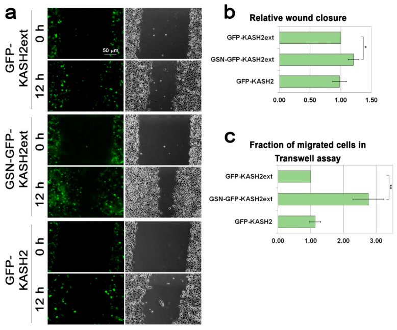
Figure 5.
Interference with the perinuclear actin rim accelerates cell migration rate. (a) Interference with perinuclear actin filaments accelerates the migration rate of B16–F10 in the wound healing assay. The migration rate of B16–F10 cells expressing GFP-KASH2ext, GSN-GFP-KAS2ext, or GFP-KASH2 was measured by the wound healing assay. Representative GFP and phase-contrast micrographs of the same fields at time 0 (immediately after the scratch) and 12 h later. Scale bar: 50 μm. (b) The area covered by the cells following 12 h of incubation was measured and set as 1 for the expressing GFP-KASH2ext cells. The graphs show the mean area covered in three independent experiments ± s.e. Statistical significance was evaluated by the Student’s t-test, * p < 0.05. (c) Interference with perinuclear actin filaments accelerates the migration rate of B16–F10 in the Transwell assay. The migration rate of B16–F10 cells expressing GFP-KASH2ext, GSN-GFP-KAS2ext, or GFP-KASH2 was measured by the Transwell assay. Four hours after plating the cells on top of the filters the cells were fixed, permeabilized, and stained with Hoechst reagent. In each experiment, the fraction of the transfected cells migrated to the lower side of the filter was calculated and normalized to the migration rate of GFP-KASH2ext-expressing cells. The graphs show the mean fraction of migrated cells in three independent experiments ± s.e. Statistical significance was evaluated by the Student’s t-test, ** p < 0.01.

