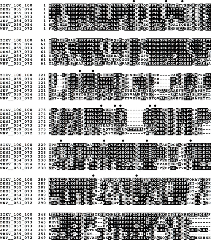
Figure 5.
Comparison of sequences (first 399 residues only) of glycoprotein E from Zika virus (ZIKV) with available X-ray structures of glycoprotein E from dengue virus type 2 (DEN2, PDB entry 1OKE), dengue virus type 2 (DEN2, PDB entry 1TG8), dengue virus type 3 (DEN3, PDB entry 1UZG), Japanese encephalitis virus (JEV, PDB entry 3P54), tick-borne encephalitis virus (TBEV, PDB entry 1SVB), and West Nile virus (WNV, PDB entry 2HG0). Multisequence alignments and this figure were generated using Clustal Omega (www.ebi.ac.uk) and Boxshade (embnet.vital-it.ch) servers. Filled circles represent residues within 5 Å of BOG based on PDB entry 1OKE (DEN2, sequence 2).

