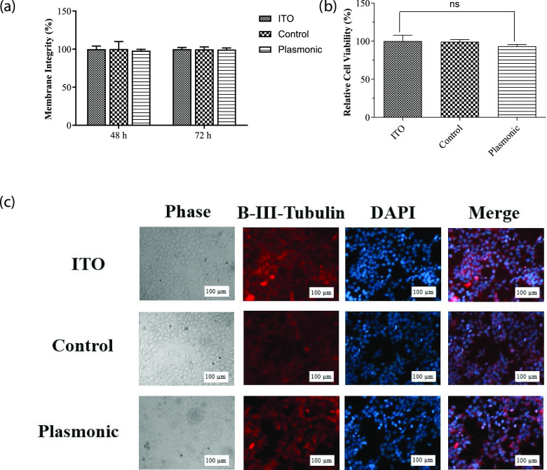
Figure 4.
(a) MTT assay, assessment of the effect of plasmonic biointerfaces on mitochondrial activity of SHSY-5Y cells. Cell viability on biointerfaces was presented relative to the ITO control. Results are presented in a column graph plotting the mean with standard error of the mean (SEM). Experiments were performed with at least three technical replicates and repeated three times (n = 3). An unpaired two-tailed t-test was performed to determine the level of significance. *p < 0.05 was considered as statistically significant; nonsignificant differences are presented as “ns.” (b) LDH leakage assay, assessment of membrane integrity of the cells grown on biointerfaces. Experiments were performed with at least three technical replicates and repeated three times (n = 3). (c) Immunofluorescence imaging, the effect of biointerfaces on the morphology of SHSY-5Y cells. The morphology of the cells grown on biointerfaces was visualized by a fluorescence microscope after β-III tubulin immunolabeling and DAPI staining (scale bar, 100 μm).

