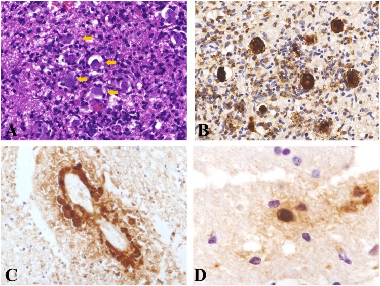
Figure 6.
Histopathology of Balamuthia mandrillaris encephalitis. A: The amebas were scattered in the brain tissue (arrow). B: Immunohistochemical staining revealed the presence of amebas. C: Immunohistochemical staining revealed a perivascular proliferation of amebas. D: Immunohistochemical staining revealed a cyst in the brain tissue.

