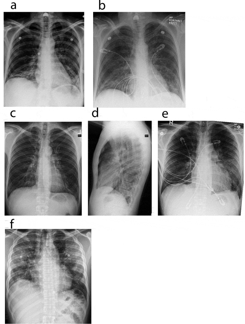
Figure 1.

Chest X ray. Chest radiographs depicting a spectrum of EVALI. (a) and (b) Chest radiographs of case 1. (a) (Day 1 of admission, pre-treatment), revealed bi-basilar haziness whereas (b) (steroid day 2), demonstrated mild improvement of left lower lobe infiltrates. (c–e) Chest radiographs of case 2. (c, d) (Day 1 of admission, pre-treatment), revealed minimal infiltrates bilaterally. Punctate density of the left upper chest is suggestive of a calcified granuloma (c), with lateral view (d) showing minimal infiltrate. (e) (steroid day 3), demonstrates clinical improvement in bilateral infiltrates. (f) Chest radiograph of case 3. (f) (Day 1 of admission, pre-treatment), revealed patchy infiltrates bilaterally
