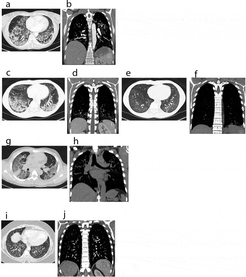
Figure 2.

CT scans: computerized tomography of chest, showing a spectrum of EVALI in axial and coronal views. (a, b) Chest CT images of case 1 on admission, pre-treatment. (a) revealed bilateral ground glass opacities and interstitial infiltrates. (b) revealed prominent right hilar lymph nodes and an enlarged intrathoracic node. (c–f) Chest CT images of case 2. (c, d) (on admission, pre-treatment) demonstrating bilateral ground glass infiltrates. (e, f) (steroid day 4) showing significant improvement of lung infiltrates bilaterally. (g, h) Chest CT images of case 3 on admission, pre-treatment. (g) demonstrating diffuse patchy ground-glass opacities bilaterally. (h) showing mediastinal lymphadenopathy and interstitial infiltrates bilaterally. (i, j) Chest CT images of an asymptomatic patient with vaping history. CT images showed mild diffuse ground-glass opacities bilaterally
