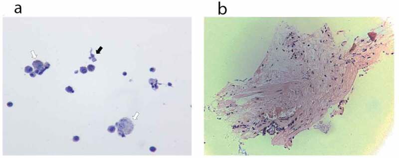
Figure 3.

Histopathology. (a) Cytology from bronchoalveolar lavage fluid of case 1. The hollow arrows point to few non-vacuolated foamy macrophages, without droplet deposition. The black arrow points to one bronchial cell. (b) Pathology from lung biopsy of case 1. Minimal tissue. There is no alveolar damage observed
