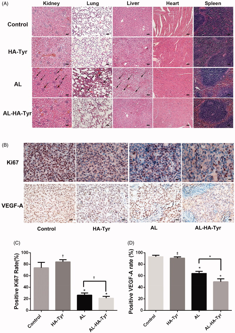
Figure 6.
Histopathology and immunohistochemistry of organs in Lewis lung cancer cell tumor-bearing mice. (A) Toxicity was evaluated by HE staining of visceral tissues. Atrophy of renal corpuscles, proliferation of alveolar cells, and ballooning degeneration of hepatocytes are indicated by black arrows. Scale bar = 50 μm. (B) Representative immunohistochemical images show Ki-67 and VEGF-A expression in tumor tissues. Scale bar = 20 μm. (C, D) Ratios (%) of Ki-67-positive cells and VEGF-A-positive cells in each group. Data are expressed as the means ± SD. †p<.05, *p<.01, ‡p>.05, HA–Tyr, AL, and AL–HA–Tyr vs. control; AL–HA–Tyr vs. AL.

