. 2020 Oct 13;9:e57117. doi: 10.7554/eLife.57117

Figure 2. Meiotic prophase is defective in Hells cKO spermatocytes.

(A) Detection of PRDM9, HELLS and SYCP3 expression in nuclear fractions of testes from Hells CTRL (Hellsfl/+ and Hellsfl/-) and Hells cKO mice at 22 dpp. Hells alleles are presented in Figure 2—figure supplement 1. HELLS and PRDM9 expression are presented in Figure 2—figure supplement 2. (B) Representative spreads of early zygotene spermatocyte nuclei from synchronized testes from Hells CTRL and Hells cKO mice after staining for DNA (DAPI, white or blue), SYCP3 (white or red) and HELLS (white or green) (top panels) or PRDM9 (white or green) (bottom panels). Anti-HELLS antibody from rabbit was used for these staining. Scale bar, 10 μm. HELLS and PRDM9 detection kinetics are presented in Figure 2—figure supplements 3 and 5. (C) Periodic acid-Schiff (PAS) staining of testis sections from 40 dpp Hells CTRL and Hells cKO mice. To visualize the glycoproteins/acrosomes (violet) and nuclei (blue), testis sections were stained with PAS and counterstained with hematoxylin. Hells CTRL testis sections (left panel) show normal spermatogenesis with well-organized stages of germ cell development, round spermatids with PAS-positive normal acrosomal caps, elongating and elongated spermatids. Hells cKO testis sections (right panel) show defective spermatogenesis with only few elongated spermatids (black arrow). Scale bar, 50 μm. (D) Proportions of seminiferous tubules without and with spermatids (mean ± SD) in testis sections from Hells CTRL and Hells cKO mice at 40 dpp. n = 4 testis sections from two mice. Data are available in Figure 2—source data 1. (E) Apoptosis detected by TUNEL assay in Hells CTRL and Hells cKO testes at 40 dpp. n = 2 testis sections from one mouse. TUNEL-positive cells are shown in Figure 2—figure supplement 4. Data are available in Figure 2—source data 1. (F) SYCP3 (red) and SYCP1 (green) staining of pachytene (Hells CTRL) and pachytene-like (Hells cKO) spermatocyte nuclei from 40 dpp mice. Arrowheads, unsynapsed chromosomes. White arrow, non-homologous synapsis. Blue arrows, sex chromosomes. Scale bar, 10 μm. (G) Representative spreads of early zygotene and pachytene or pachytene-like spermatocyte nuclei from 40 dpp Hells CTRL and Hells cKO mice, respectively, after staining for SYCP3 (white or red), DMC1 (white or green) and γH2AFX (white or blue). Scale bar, 10 μm.

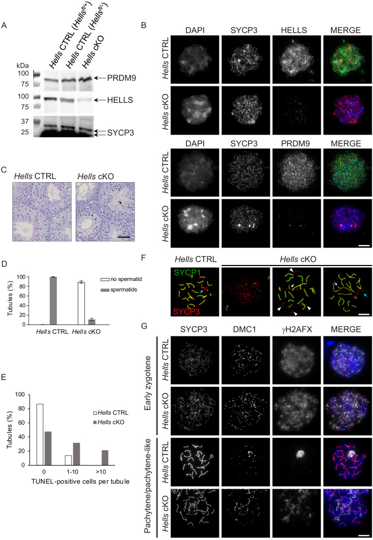
Figure 2—source data 1. Quantification of spermatid and TUNEL-positive sections.

Figure 2.

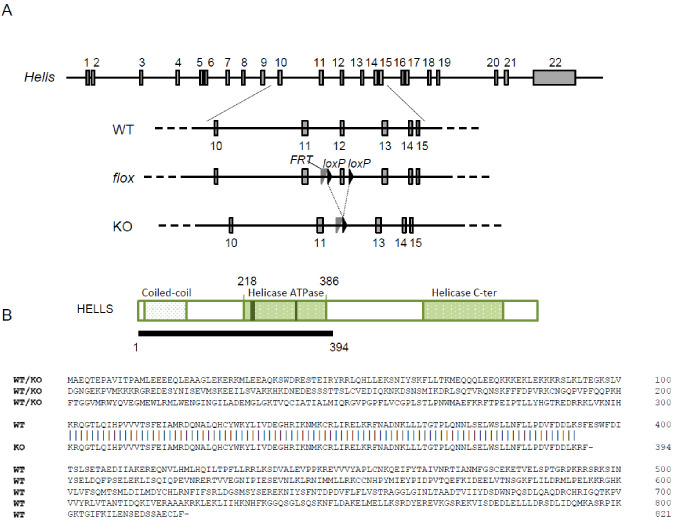
Figure 2—figure supplement 1. The Hells cKO allele.

Figure 2—figure supplement 1.

(A) Schematic diagram of the Hells alleles used in this study. (B) Schematic diagram (top) and predicted sequence (bottom) of wild-type (from WT or floxed allele) and mutant (KO) HELLS proteins.
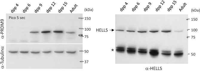
Figure 2—figure supplement 2. PRDM9 and HELLS protein levels during the first wave of spermatogenesis in wild-type mice.

Figure 2—figure supplement 2.

Testis extracts from RJ2 mice at the indicated days post-partum (dpp) and from adult were analyzed by western blotting. The same amount of extracts was loaded in parallel on two different gels. One membrane (left) was probed with anti-PRDM9 and anti-tubulin α antibodies, the other membrane (right) with an anti-HELLS antibody. The star on the right panel indicates non-specific bands.
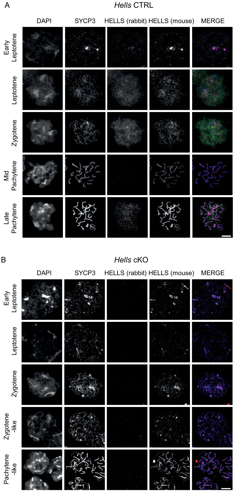
Figure 2—figure supplement 3. HELLS detection in Hells CTRL and Hells cKO spermatocytes Representative spreads of spermatocyte nuclei from Hells CTRL.

Figure 2—figure supplement 3.

(A) And Hells cKO (B) Mice after staining for DNA (DAPI, white), SYCP3(white or blue), and HELLS using a rabbit (white or green) and a mouse (white or red) antibody. Scale bar, 10 μm. Both rabbit and mouse anti-HELLS antibodies display a nucleus-wide, punctate staining in leptotene and zygotene stages, attributable to HELLS. In addition to this specific signal, both antibodies showed also a different non-specific signal. The mouse antibody showed a colocalization with SYCP3 at every stage, that was not seen with the rabbit antibody and is therefore most likely not attributable to HELLS. The rabbit antibody showed a nucleus-wide staining in late pachytene and diplotene stage nuclei, not seen with the mouse antibody, likely due to cross-reaction.
Figure 2—figure supplement 4. TUNEL-positive cells detected in testis sections of control and Hells cKO mice.

Figure 2—figure supplement 4.

Sections from 40dpp Hells CTRL and Hells cKO mice were stained for DNA (DAPI, purple) and TUNEL (green). Asterisks mark tubules containing round spermatids. Scale bar, 10 μm.
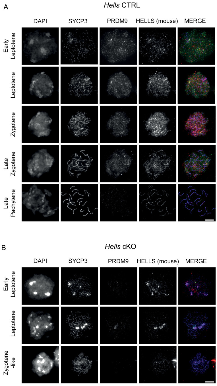
Figure 2—figure supplement 5. HELLS and PRDM9 detection in Hells CTRL and Hells cKO spermatocytes.

Figure 2—figure supplement 5.

Representative spreads of spermatocyte nuclei from Hells CTRL (A) and Hells cKO (B) mice after staining for DNA (DAPI, white), SYCP3 (white or blue), PRDM9 (white or green) and HELLS (white or red). Scale bar, 10 μm. In addition to bona fides HELLS staining, the mouse anti-HELLS antibody displays additional, non-specific staining marking the chromosome axes, as described in the legend to Figure 2—figure supplement 3.