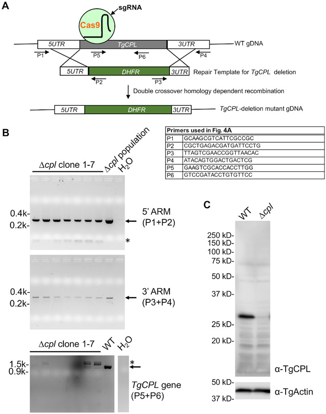
Figure 4: PCR and immunoblotting confirmation of TgCPL-deficient parasites.
(A) A schematic diagram depicting the general strategies of TgCPL-deletion in Toxoplasma and PCR-based screening of the correct TgCPL knockout clones. The primers used for the screening are labeled. (B) PCR and agarose gel electrophoresis were used to select clones containing the correct integration of the pyrimethamine resistance cassette into the TgCPL locus and loss of the TgCPL gene. The genomic DNA of the Δcpl population served as a positive control for 5'- and 3'-ARM detection, while the WT genomic DNA was used for the detection of the TgCPL gene as a positive control. Water was used instead of DNA template in the PCR reactions to serve as a negative control. The expected bands are denoted by arrows, whereas nonspecific PCR amplifications are labeled by asterisks. (C) Clone 1 identified by PCR screening was grown in tissue culture for cell lysate preparation and further immunoblotting analysis to confirm the loss of TgCPL expression in the knockout. TgActin was used as a loading control.

