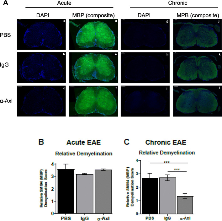
Fig. 2.
Demyelination is significantly reduced during chronic EAE in response to α-Axl antibody treatment. Representative lumbar spinal cord sections from C57Bl/6J mice treated with PBS (n = 6), IgG isotype control (n = 6), or α-Axl-activating antibody (n = 6) analyzed by IHC. A Immunofluorescent staining of the representative spinal cord sections during acute (a–f) and chronic (g–l) EAE. blue = DAPI, (d–f) composite of MBP (green) and DAPI. Quantification of MBP demyelination during acute (B) and chronic (C) EAE by scoring the criteria detailed in the “Methods” section. *p ≤ 0.05 and **p ≤ 0.01, Mann-Whitney U test. All scale bars are 200 μm

