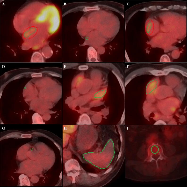
Figure 1.
Representative axial positron emission tomography/computed tomography (PET/CT) images showing examples of regions of interest (ROIs) in the left atrial wall with (A) and without (B) visual uptake, right atrial wall with (C) and without (D) visual uptake, left atrial appendage (E), right atrial appendage (F), right coronary artery (G), spleen (H), and bone marrow (I)

