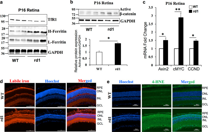
Fig. 3. Iron accumulation and enhanced Wnt signaling in the rd1 mouse model of Retinitis Pigmentosa.
a TfR1, Heavy (H-) chain ferritin, light (L-) chain ferritin, and b active β-catenin protein levels were determined by immunoblotting in WT and rd1 mouse at postnatal day 16. Blots cropped from different parts of the same gel or from different gels are separated by white space in a and b. c mRNA expression of Wnt downstream target genes Axin2, cMYC, CCND in WT, and rd1 mice retina at P16. d Representative images of retinal sections from P16 WT and rd1 mice stained for labile iron using FeRhoNox-1 fluorescent imaging probe. e 4-Hydroxynonenal staining in WT and rd1 mouse retina at P16. Scale bar is 100 μm for all the panels in d and e. Data presented as mean ± SE of three independent experiments; n = 3 mice per group; *p < 0.05; **p < 0.001.

