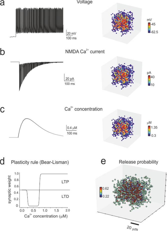
Fig. 5. Simulation of synaptic plasticity.
a–c Cellular firing caused by HFS (the same as in the experimental induction protocol) determines Ca2+ influx through synaptic NMDA receptor channels and the consequent increase in intracellular Ca2+ concentration. The membrane voltage (average voltage), NMDA current (integral of the current) and Ca2+ concentration (peak concentration) vary from cell to cell depending on the local connectivity inside the microcircuit unit, as shown in the 3D activity maps. d The Bear–Lisman rule was used to calculate the neurotransmission changes induced by the Ca2+ concentrations reached during HFS. e The mossy fiber release probability changed accordingly (plotted here over the receiving granule cells) to account for LTP or LTD expression.

