
Figure 2.
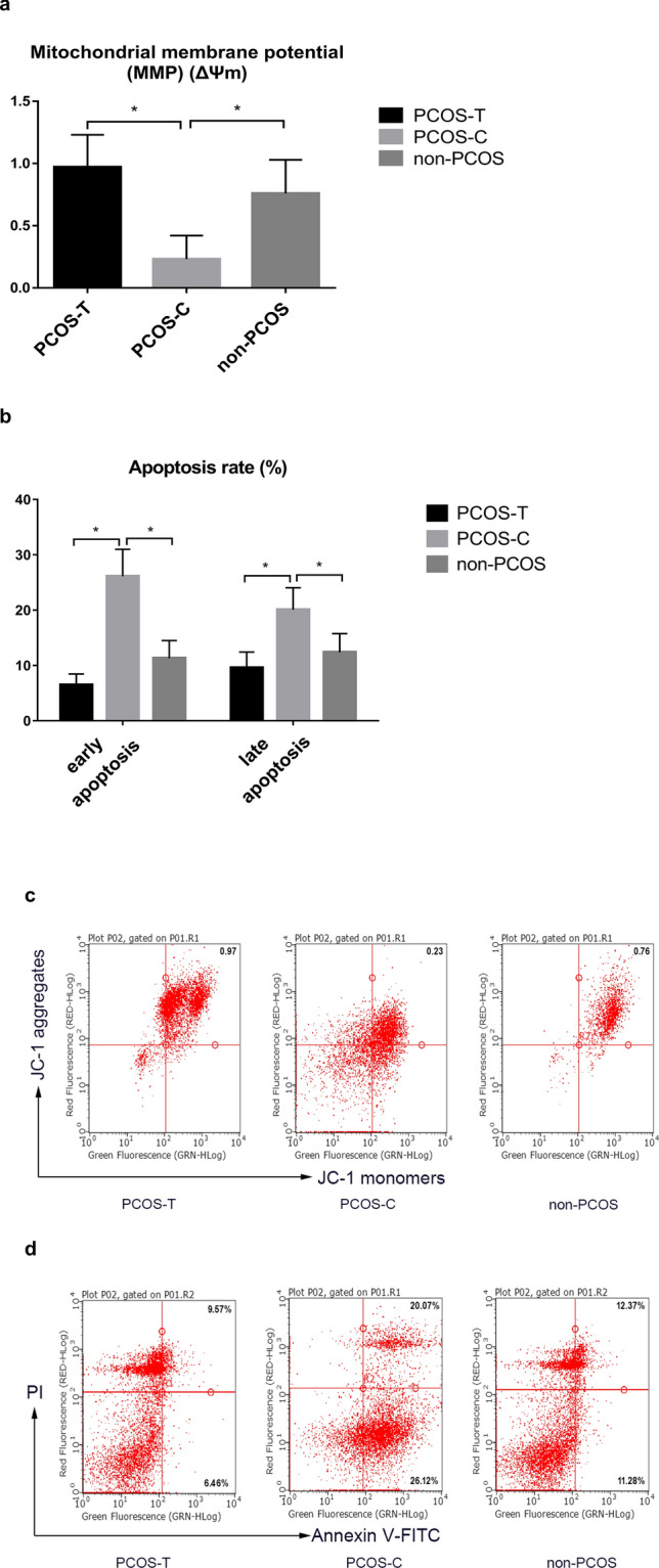
Flow cytometric dot plots of effects of GH on mitochondrial function in granulosa cells. (a, c) The MMP in GCs was lower in the PCOS-C group than in the non-PCOS control group, as shown by reduced JC-1 aggregates red fluorescence. The MMP was greater in the PCOS-T group than in the PCOS-C group (P < 0.05). (b, d) The numbers of early and late apoptotic cells were increased in the PCOS-C group compared with those in the non-PCOS and PCOS-T groups (P < 0.05). GH growth hormone, MMP mitochondrial membrane potential, PCOS polycystic ovary syndrome, PI propidium iodide, FITC fluorescein isothiocyanate. *P < 0.05 denotes statistical significance between groups.