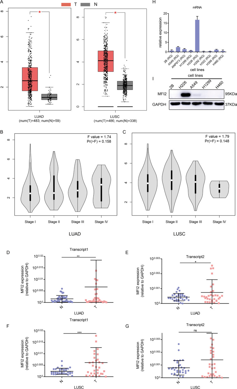
Fig. 1. MFI2 is significantly upregulated in lung cancer tissues and cell lines.
A Expression of MFI2 in GEPIA databases. The left is the expression of 483 lung adenocarcinoma tissues and 59 normal tissues, and the right side is the expression of 486 lung squamous cell carcinoma tissues and 338 normal tissues. LUAD, lung adenocarcinoma, LUSC, lung squamous cell carcinoma. B, C Expression of MFI2 in different stages of lung adenocarcinoma and lung squamous cell carcinoma. D–G The expression of two transcripts of MFI2, which were normalized to that of GAPDH, in fresh-frozen samples of Lung adenocarcinoma and lung squamous cell carcinoma, as determined by RT-qPCR. H, I Detection of MFI2 expression in common cell lines by RT-qPCR and western blot. Data are presented as the mean ± SD, n = 3. *p < 0.05, **p < 0.01, ***p < 0.001. ns means no significance.

