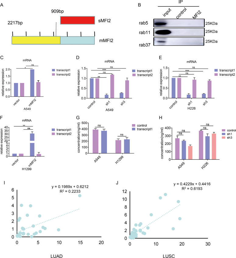
Fig. 6. mMFI2 and sMFI2 are independent of each other in source and function.
A Schematic diagram of mMFI2 and sMFI2 coding region (CDS). B Cellular lysates were extracted from A549 with overexpression of mMFI2 were subjected to immunoprecipitation with anti-mMFI2 antibody, followed by anti-rab5, anti-rab11, and anti-rab37 immunoblotting. C–H RT-PCR analysis of expression levels of mMFI2 (transcript 1) and sMFI2 (transcript 2) in cell clones A549, H1299, and H226 which had overexpression or knocking down of mMFI2. I, J Correlation analysis of expression levels of mMFI2 (transcript 1) and sMFI2 (transcript 2) expression in frozen tissues of lung adenocarcinoma and lung squamous cell carcinoma. Data are presented as the mean ± SD, n = 3. *p < 0.05, **p < 0.01, ***p < 0.001.

