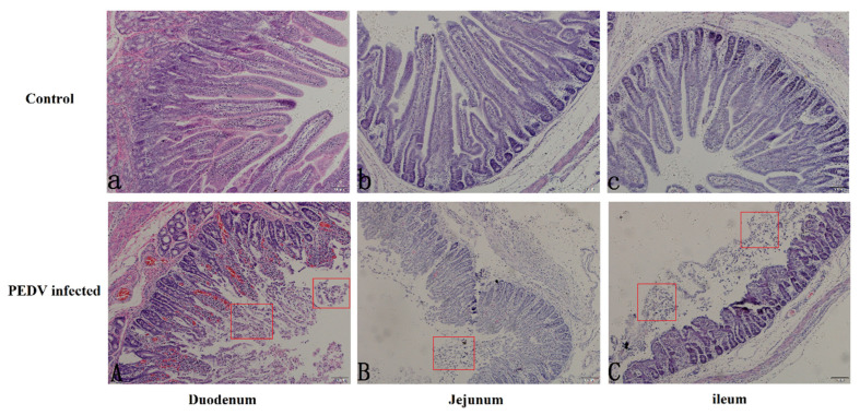
Figure 1.
The paraffin sections microscopy of three sections of the bowel in pig intestine (10×). The top group represents the duodenum (a), jejunum (b), and ileum (c) of normal piglets, and the bottom group represents the duodenum (A), jejunum (B), and ileum (C) of diarrheic piglets. The red square marks the area of intestinal villus loss.

