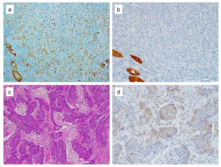
Figure 2.
MSH2 cytoplasmic expression. (a) MSH2 cytoplasmic expression in both normal epithelial glands and in medullary colorectal cancer cells. Notice the lack of nuclear MSH2 expression in neoplastic cells. Lymphocytes in between the tumor cells showed nuclear staining and served as the internal positive control. (b) Loss of EPCAM expression in the tumor cells with normal colonic glands retaining the staining. (c) H&E staining of poorly differentiated gastric carcinoma displaying a cohesive solid pattern. (d) MSH2 staining yields a cytoplasmic expression in some of the tumor cells.

