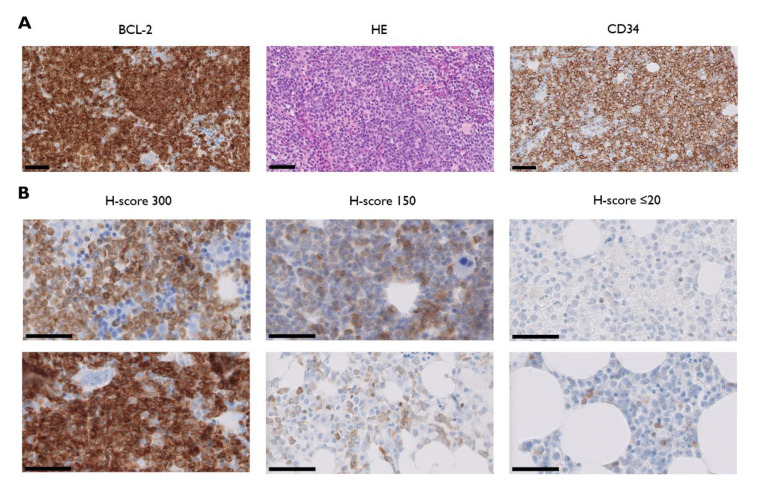
Figure 1.
(A) Light micrographs of BCL-2 (B-cell leukemia/lymphoma-2, left) and corresponding haematoxylin and eosin (HE; middle) and CD34 stained paraffin sections of trephine biopsies from one representative acute myeloid leukemia patient with a BCL-2 H-score of 300; (B) bone marrow BCL-2 immunostains from two representative patients with H-scores of 300 (left panel), 150 (middle panel), and ≤20 (right panel). Thick black line = 50 µm.

