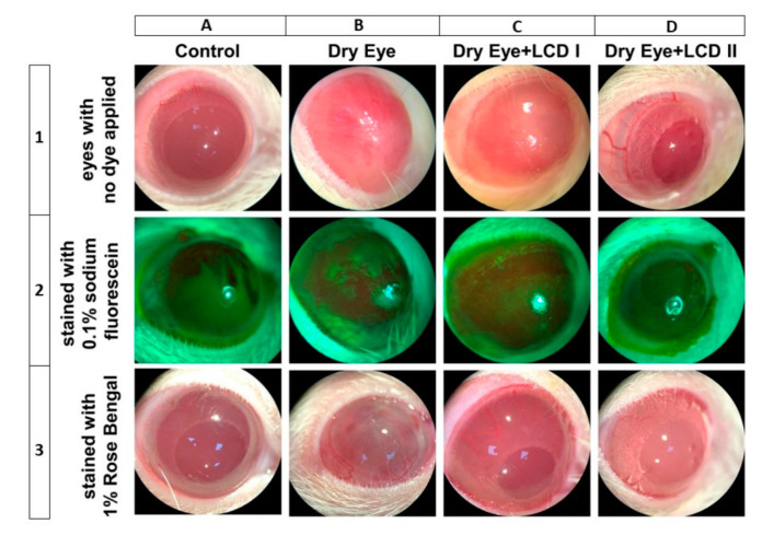
Figure 1.
Effect of the combination of lutein/zeaxanthin, curcumin, and vitamin D (LCD) on Slit-lamp microscopic images of eyes in benzalkonium chloride (BAC) induced dry eye syndrome in rats. Control: No treatment; Dry Eye: BAC-induced dry eye syndrome; LCD I; Dry Eye + LCD I: BAC-induced dry eye syndrome plus LCD I (100 mg/kg body weight); Dry Eye + LCD II: BAC-induced dry eye syndrome plus LCD II (200 mg/kg body weight). Each representative vertical column contains the images of eyes from different groups (n = 7 per group); Horizontal rows respectively from up to down contain images of eyes with no dye applied (upper row, 1), stained with 0.1% sodium fluorescein (middle row, 2) and stained with 1% Rose Bengal (lower row, 3). A normal ocular surface of the rat in the control group is shown in A1. The cornea is transparent and the details of the iris and pupil can be seen behind the cornea. No sign of inflammation and pathologic staining with sodium fluorescein (A2) and Rose Bengal (A3) was observed on the ocular surface of rat eyes belongs to the control group. Slit-lamp microscopy shows us more than 2 mm ciliary hyperemia and vascularized cornea of rat induced by BAC (B1). Iris and pupil are invisible due to the scar and vascularization. Diffuse staining with plaques (B2) and confluent spots (B3) are observed in stained corneas by sodium fluorescein and Rose Bengal, respectively. The dose-dependent positive effect of LCD is seen in the images C1 and D1. Scar formation and vascularization of the cornea were recovered partly and almost fully in LCD I (C1) and LCD II (D1) groups, respectively. Iris and pupil details can be distinguished in C1 and more clearly seen in the D1. Similarly, lesser staining of the ocular surface was observed for both sodium fluorescein and Rose Bengal dye in LCD I (C2-3) and LCD II (D2-3) groups.

