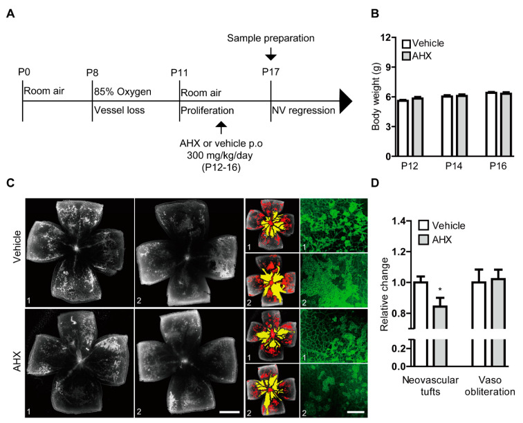
Figure 4.
Suppression of neovascularization by oral administration of AHX. (A) The schematic illustration shows the murine OIR model procedure and the oral administration of the AHX or vehicle to mice. (B) Quantitative analyses for body weights of the mice (n = 5–6 per group) showed that the average body weights of the AHX-administered group (three male and three female pups) did not significantly differ from those of the vehicle group (three male and two female pups) during the administration period. (C) Representative images of retinal vascular whole mount (vehicle 1, 2 and AHX 1, 2) staining with isolectin B4 (neovascular tufts: red and vaso-obliteration: yellow), and higher-magnification images (green) for neovascular tufts. Scale bars are 1000 and 200 µm, respectively. (D) Quantitative analyses showed that areas of neovascular tufts (red) were suppressed in the AHX-administered group (n = 6) in comparison with those in the vehicle-administered group (n = 5), while no significant difference in vaso-obliteration (yellow) was observed between the groups. * p < 0.05. Bar graphs were presented as mean with ± standard error of the mean. The data were analyzed using a two-tailed Student’s t-test.

