Figure 2.
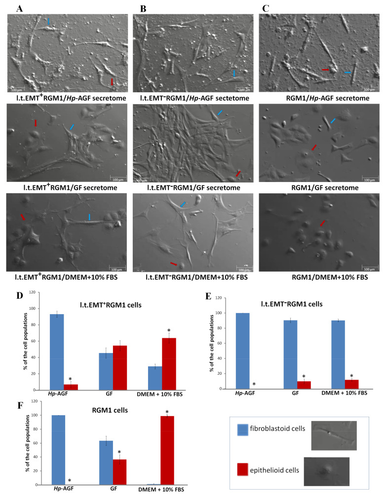
The phenotypical plasticity of l.t.EMT+RGM1 cells. Nomarski contrast of the stability of phenotype of (A) l.t.EMT+RGM1 cells placed in Hp-AGF secretome, GF secretome, and DMEM + 10% FBS, respectively; (B) l.t.EMT−RGM1 cells placed for 24 h in Hp-AGF secretome, GF secretome, and DMEM + 10% FBS, respectively; (C) and of RGM1 cells placed for 24 h in Hp-AGF secretome, GF secretome, and DMEM + 10% FBS, respectively. L.t.EMT+RGM1 cells were characterized by plastic phenotype which depended on the type of the medium suggesting EMT/MET switch ability. L.t.EMT−RGM1 cells cultured for 24 h in DMEM + 10% FBS possessed stable, fibroblastoid-like phenotype in all tested media. Histogram analysis of morphological cell diversity in (D) l.t.EMT+RGM1 cells placed in Hp-AGF secretome, GF secretome, and DMEM + 10% FBS, respectively; (E) l.t.EMT−RGM1 cells placed for 24 h in Hp-AGF secretome, GF secretome, and DMEM + 10% FBS, respectively; (F) and of RGM1 cells placed for 24 h in Hp-AGF secretome, GF secretome, and DMEM + 10% FBS, respectively. The cells were counted according to cell morphology (non-polarized epithelioid cells vs rear-front polarized fibroblastoid cells). Blue arrows: sample fibroblastoid cells, red arrows: sample epithelioid cells. Results are mean ± SEM of three to six different independent experimental repeats. Asterisk indicates a significant difference between epithelial and fibroblastoid populations (p < 0.05).

