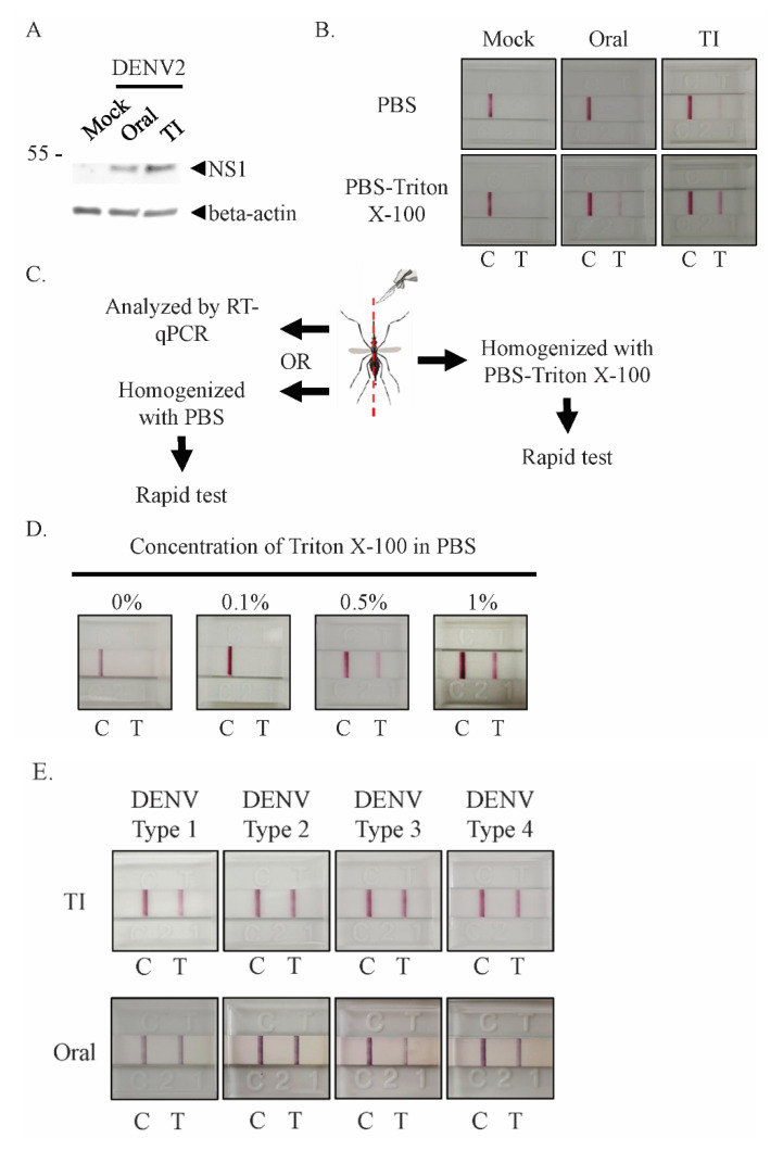
Figure 2.
NS1 can be detected in lysate from mosquito bodies when Triton X-100-PBS lysis buffer is used. (A) Mosquitoes were infected with DENV2 orally (oral) or via thorax injection (TI). After seven days of incubation, infected mosquitoes were homogenized with lysis buffer. Soluble lysates were separated with SDS-PAGE and immunoblotting was performed with anti-NS1 and beta-actin antibodies. (B–E) Mosquitoes were orally (oral) or intrathoracically infected (TI) with DENV2 and incubated for seven days. (B) Incubated mosquitoes were homogenized with PBS or 1% Triton X-100-PBS buffer; both lysates were then tested using the NS1 rapid test. (C) To confirm the infection status of orally infected mosquitoes, mosquitoes were longitudinally and symmetrically bisected for homogenization in different lysis buffers or for analysis with different methods. (D) The Triton X-100-PBS lysis buffer dose-dependent assay for orally infected mosquitoes. Half of each infected mosquito was homogenized with 0.1–1% Triton X-100-PBS buffer and tested using the NS1 rapid test strip. (E) Mosquitoes were infected with one of the four DENV serotypes via thorax injection or oral infection, before being homogenized with 1% Triton X-100-PBS and tested using the rapid test strip. At least 5 individual mosquitoes were tested per sample. The DENV titer from serotype1 to 4 of TI infected mosquito was 2.2 × 10⁴, 4.0 × 10⁴, 2.9 × 10⁴, and 2.3 × 10⁴, respectively. The virus titer from serotype1 to 4 of orally infected mosquito was 8.9 × 10³, 1.1 × 10⁴, 6.8 × 10³, and 8.0 × 10³, respectively. All control (C) and positive (T) signals were observed within 20 min.

