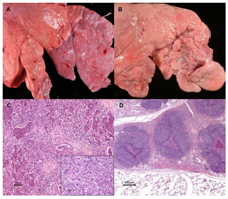
Figure 1.
Chronic catarrhal bronchopneumonia. (A). Demarcated area of cranioventral lung consolidation with purple tonality, firm consistency and no increase of volume. Mucopurulent content and bronchiectasis were observed (inset). (B). Lesion showing recovery features such as coalescing small purple foci, consistent mainly with atelectasis surrounded with lobules with variable degrees of emphysema. (C). Haematoxylin-eosin staining (H-E). Histologically, neutrophils and, less in number, macrophages, occupy the bronchioalveolar lumen; foci of newly epithelization (inset) were observed. (D). Areas corresponding with atelectasis associated with occlusion of the bronchiolar lumen by inflammatory exudate and the peripheric lymphoid hyperplasia.

