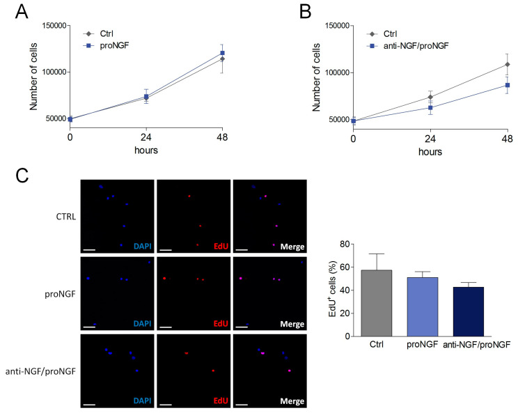
Figure 2.
Effect of proNGF administration and anti-NGF/proNGF addition on SCDM proliferation. (A) SCDMs were cultured in a proliferation medium and treated with proNGF (100 ng/mL) up to 48 h. Cells were counted at both 24 and 48 h to evaluate cell growth. (B) SCDM cell growth was evaluated as in A, in both control cells and SCDMs treated with neutralizing anti-NGF/proNGF antibody (500 ng/mL) for 48 h. (C) EdU staining and the ratio of EdU+ cells from proliferating SCDMs, kept in a proliferation medium in the presence or not of proNGF (100 ng/mL) or anti-NGF/proNGF (500 ng/mL) for 48 h. DAPI was used to counterstain the nuclei. Three images for each experimental group were captured under a fluorescence microscope, and then analyzed to evaluate the percentage of EdU+ cells. All the experiments consist of at least three biological replicates. Scale bar: 50 μm. Values are expressed as the mean ± SD. For cell proliferation, statistical analysis was performed by using two-way ANOVA, whereas the ratio of EdU+ cells was analyzed by one-way ANOVA followed by Tukey’s post hoc test.

