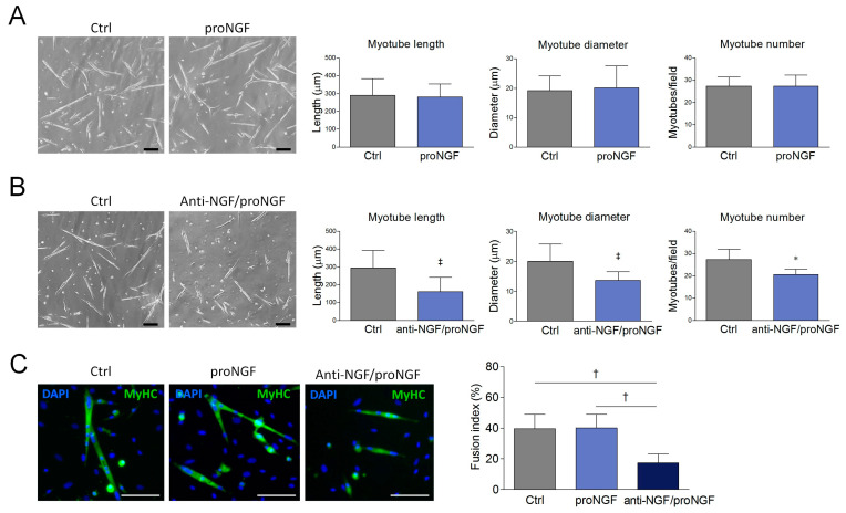
Figure 3.
Effect of proNGF administration and anti-NGF/proNGF addition on SCDM differentiation. (A) SCDMs were allowed to differentiate into myotubes in the differentiation medium (DM), in presence or not of proNGF (100 ng/mL) for 72 h. Cells were observed with bright field microscopy and three images for every single experiment were captured. (B) SCDMs were treated with anti-NGF/proNGF (500 ng/mL) and induced to differentiate in DM for 72 h. Three images for each experimental group were captured under a brightfield microscope, and then analyzed to evaluate myotube length, myotube diameter and myotube number. (C) SCDMs were induced to differentiate in the presence of proNGF (100 ng/mL) or anti-NGF/proNGF (500 ng/mL) for 72 h. Once differentiation was completed, the cells were fixed in paraformaldehyde (PFA) 4% and incubated overnight with anti-MyHC (green) to evaluate the fusion index by immunofluorescence. DAPI (blue) was used to counterstain the nuclei. Fusion index was calculated as the percentage of nuclei incorporated into myotubes on the total nuclei present in each field. All the experiments consisted of at least three biological replicates. Scale bars: 100 μm. Values are expressed as the mean ± SD. Statistical analysis was performed by using the Student’s t test. * p < 0.05; † p < 0.01; ‡ p < 0.001.

