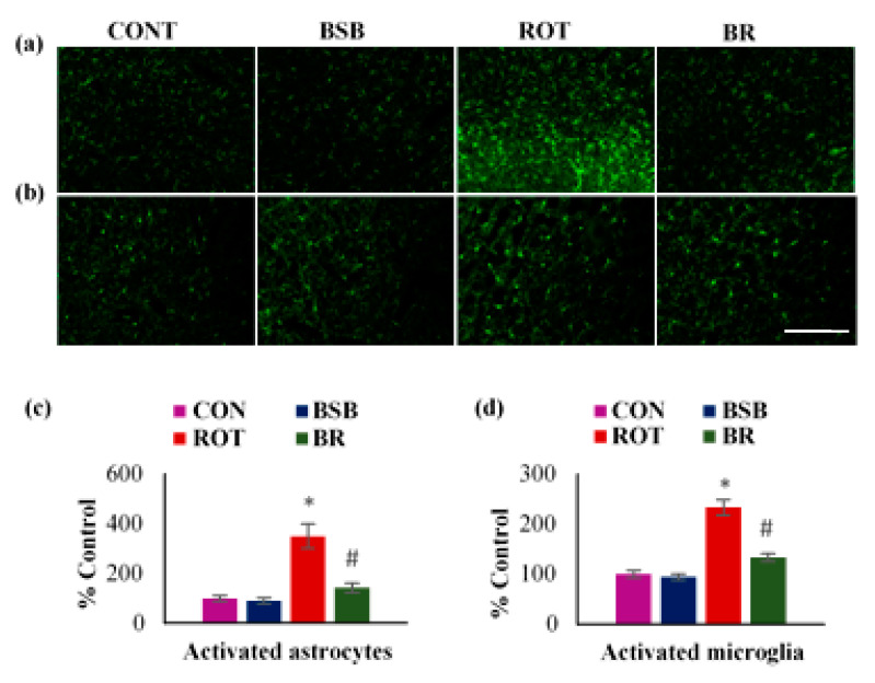
Figure 5.
The immunofluorescence expression of GFAP and Iba-1 in striatum was determined. A profound expression of GFAP-positive astrocytes (a), and Iba-1-positive microglia (b), was observed in the ROT-injected rats in comparison with vehicle treated control (CON) rats. Interestingly, BSB treatment to ROT injected rats showed decreased expression of GFAP and Iba-1 compared to ROT injected rats (scale bar = 200 µm). The quantitative representation of the result of activated astrocytes and microglia in the striatum is shown (c,d). Each group contained three rats and the data were expressed as percent mean ± SEM. * p < 0.05 CON vs ROT; # p < 0.05 ROT vs. BR (One-way ANOVA followed by Tukey’s test).

