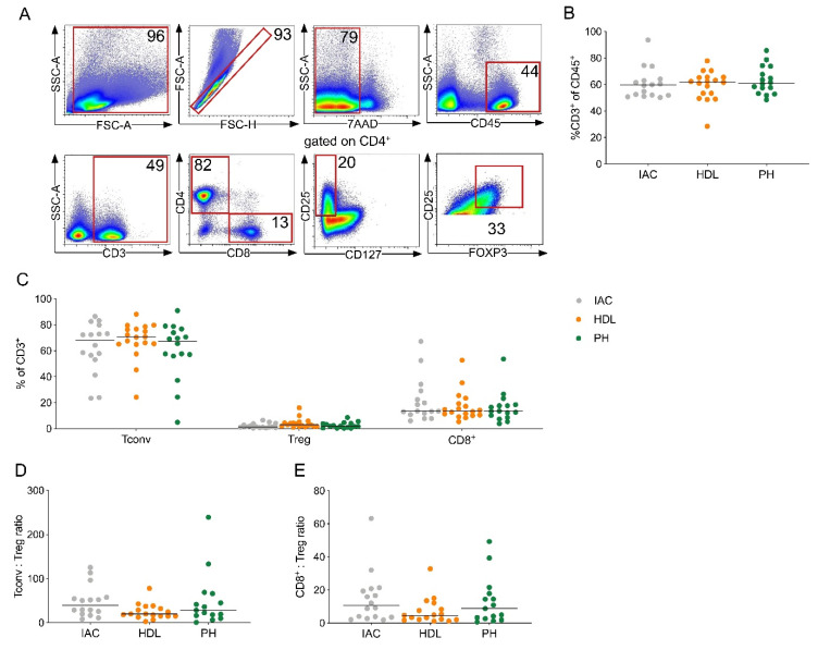
Figure 1.
CD4+ T cells are the predominant T cell subset in pancreatic ductal adenocarcinoma (PDAC)-draining lymph nodes. (A) Representative flow cytometric gating strategy for the identification of T cells. Number indicates percentage of population per gate. SSC, side scatter; FSC, forward scatter. (B) Quantification of CD3+ T cells among all leucocytes (CD45+). (C) CD4+ Tconv cells (Tconv; CD3+CD4+’not Treg’), regulatory T cells (Treg; CD3+CD4+CD8-CD25+CD127-FOXP3+) and CD8+ T cells (CD8+; CD3+CD4-CD8+) as a percentage of CD3+ T cells in lymph nodes of the indicated location from patients with PDAC. (D) Ratio of CD4+ Tconv to Treg and (E) CD8+ T cells to Treg in lymph nodes. IAC, interaortocaval: lymph node around the abdominal aorta; HDL, hepatoduodenal ligament: lymph node along the hepatic artery and bile duct; PH, pancreatic head: lymph node from the posterior aspect of the pancreatic head. Each point represents data from one patient. Data, median. One-way ANOVA.

