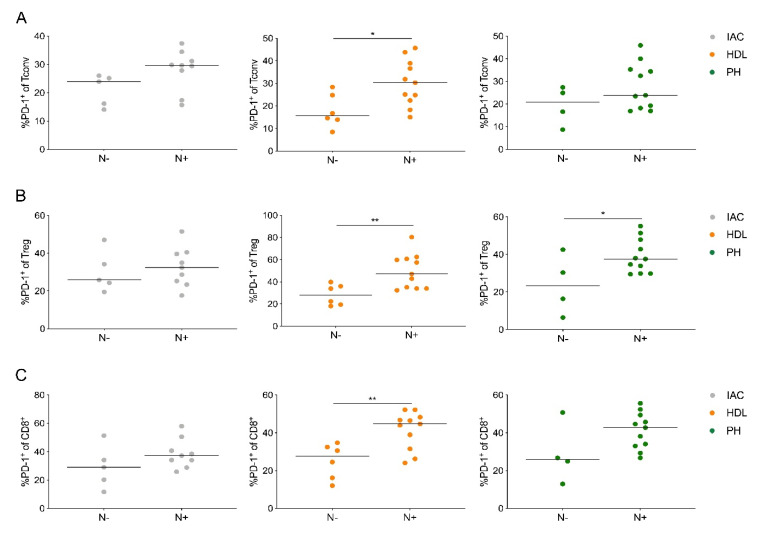
Figure 5.
PD-1-expressing lymph node T cells are associated with node-positive PDAC. (A) Quantification of the expression of PD-1 on CD4+ Tconv cells (Tconv), (B) regulatory T cells (Treg) and (C) CD8+ T cells (CD8+) based on nodal stage (N-, negative; N+, positive). IAC, interaortocaval; HDL, hepatoduodenal ligament; PH, pancreatic head. Each point represents data from one patient. Data, median. Unpaired t-test. * p < 0.05, ** p ≤ 0.01.

