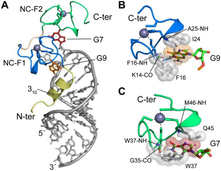
Figure 15.
NMR structure of HIV-1NL4-3 NC bound to the GGAG loop region of the Ψ-hairpin stem-loop [109]. (A) Overall view showing the relative orientation of the RNA (gray) relative to the 310-helix (yellow), N-terminal zinc knuckle (F1, blue), and C-terminal zinc knuckle (F2, green). Guanosines G7 and G9 are shown in red and orange, respectively. (B) Interactions between G9 and NC-F1. Hydrogen bonds are depicted as yellow dash lines and hydrophobic side chains shown as spheres. (C) Interactions between G7 and NC-F2.

