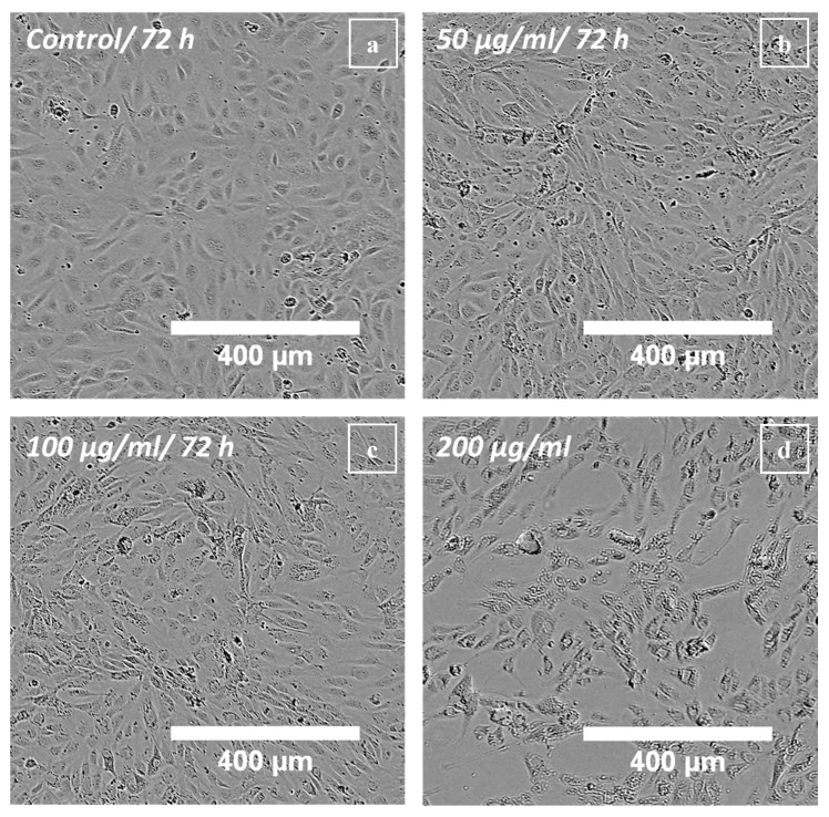
Figure 4.
Live-cell microscopy of HUVECs treated with polyR-Fe3O4: Phase contrast images of HUVECs were taken after 72 h post particle addition. (a) Untreated control HUVECs, (b) HUVECs treated with 50 µg mL−1, (c) HUVECs treated with 100 µg mL−1 and (d) HUVECs treated with 200 µg mL−1 polyR-Fe3O4. Representative images of n = 3 experiments and hexaplicate samples are shown.

