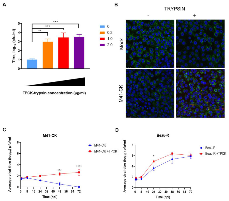
Figure 1.
Trypsin enhances infectious bronchitis virus (IBV) replication in Vero cells. (A) Vero cells were infected with M41-chicken kidney (CK) (MOI = 1) diluted in 1XBES medium containing increasing concentrations of trypsin treated with l-(tosylamido-2-phenyl) ethyl chloromethyl ketone (TPCK-treated trypsin) from 0–2.0 µg/mL. Supernatant was harvested at 24 hpi and titrated on CK cells in triplicate. Data are representative of three biological replicates. Data were analysed by One-Way ANOVA followed by Tukey’s test for multiple comparisons and statistical differences from the untreated (0 µg/mL TPCK) values are indicated. ** indicates p < 0.01, *** indicates p < 0.001, **** indicates p < 0.0001. (B) Confocal images of Vero cells infected with M41-CK in the presence of trypsin (1.0 µg/mL) compared to mock infected and untreated cells. Cells were stained with monoclonal antibodies against dsRNA (Red) and α-tubulin (Green). Nuclei were stained with DAPI (Blue). White scale bars indicate 25µm. Replication kinetics of M41-CK (C) and Beau-R (D) in the presence of trypsin (1.0 µg/mL) were assessed in Vero cells. Cells were infected with IBV at MOI of 0.01. Supernatant was harvested at 1, 12, 24, 48 and 72 hpi and titrated on CK cells in triplicate. Average viral titres of at least three biological replicates are displayed with SEM. Statistical differences were calculated by Two-Way ANOVA comparing IBV strains and treatments.

