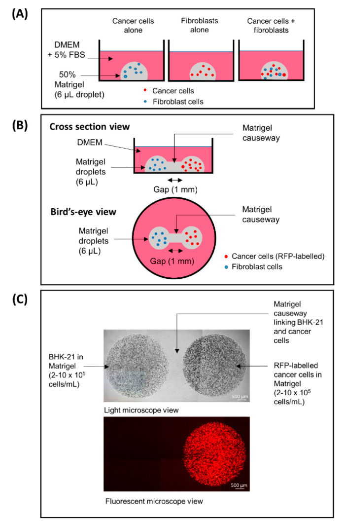
Figure 1.
(A) A conventional 3D co-culture model, (B,C) a “3D dumbbell model” invented in this study for the visualization of cancer-fibroblast cell interaction. (A) CaKi-1 and BHK-21 fibroblast cells embedded in 50% Matrigel suspension (6 μL/droplet; containing 5 × 10⁵ cells/mL) as independent or co-culture droplets. (B) Caki-1 cells and BHK-21 fibroblasts in 50% Matrigel suspension (6 μL/droplet; 5 × 10⁵ cells/mL) droplets bridged by a Matrigel causeway that allows for cell migration and interaction. (C) Note that the cancer cells are labelled with mCherry red fluorescent protein (RFP) and thus appeared red under a fluorescent microscope. Brightfield and fluorescent views of the same cells under a 40× low power objective lens.

