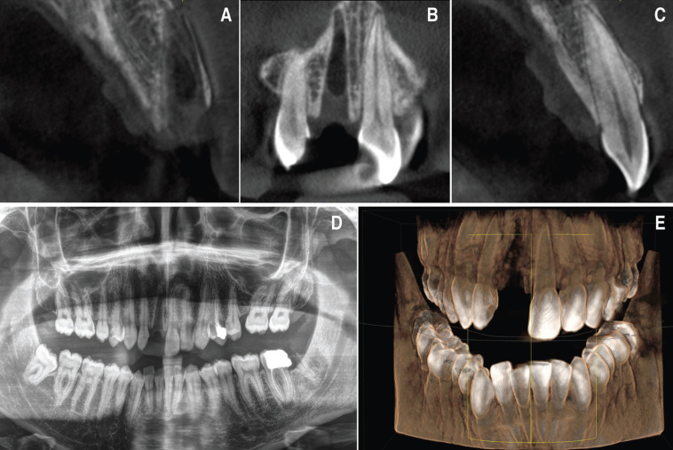
Figure 2.
Pre-treatment radiographic study. A) Axial tomographic cut of the socket after the avulsion of the right upper incisor. B) Coronal view of the socket after the avulsion. C) Axial tomographic cut of the upper left incisor. D) Initial orthopantomography after tooth avulsion. E) 3D reconstruction of the patient’s bone jaws and teeth following dental trauma.

