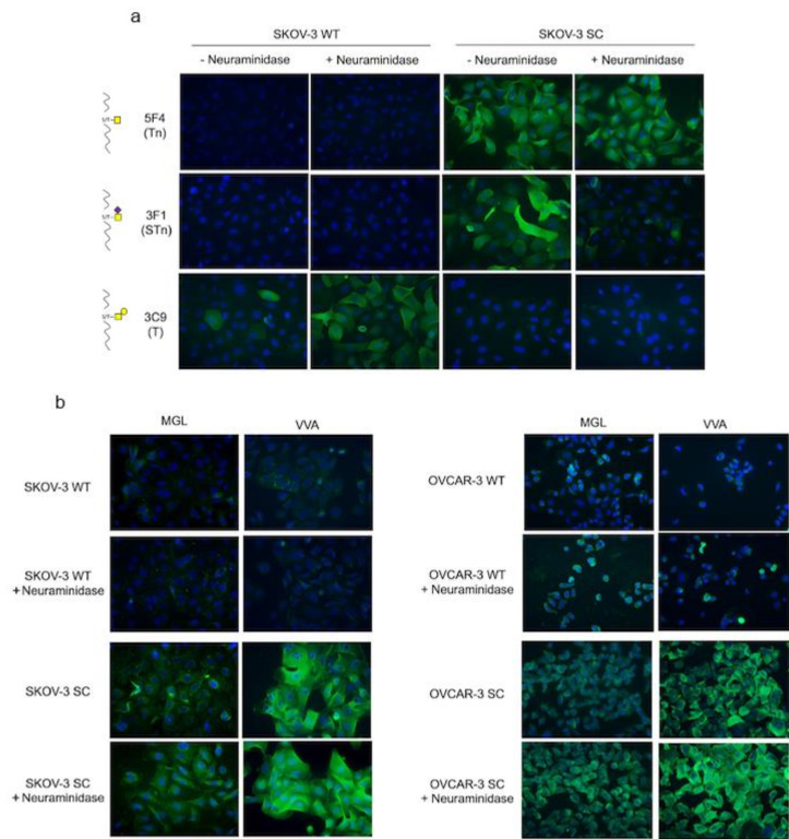
Figure 2.
Glycoprofiling and lectin binding of SKOV-3 and OVCAR-3 ovarian cancer cell lines. (a) Immunofluorescence of wild-type (WT) and SimpleCell (SC) modified SKOV-3 cell line with and without neuraminidase treatment stained with anti-Tn mAb 5F4, anti-STn, mAb 3F1, and anti-T mAb 3C9. (b) Fluorescence microscopy of WT and SC SKOV-3 and OVCAR-3 with and without neuraminidase treatment, stained with rhMGL or VVA lectin (magnification 20×).

