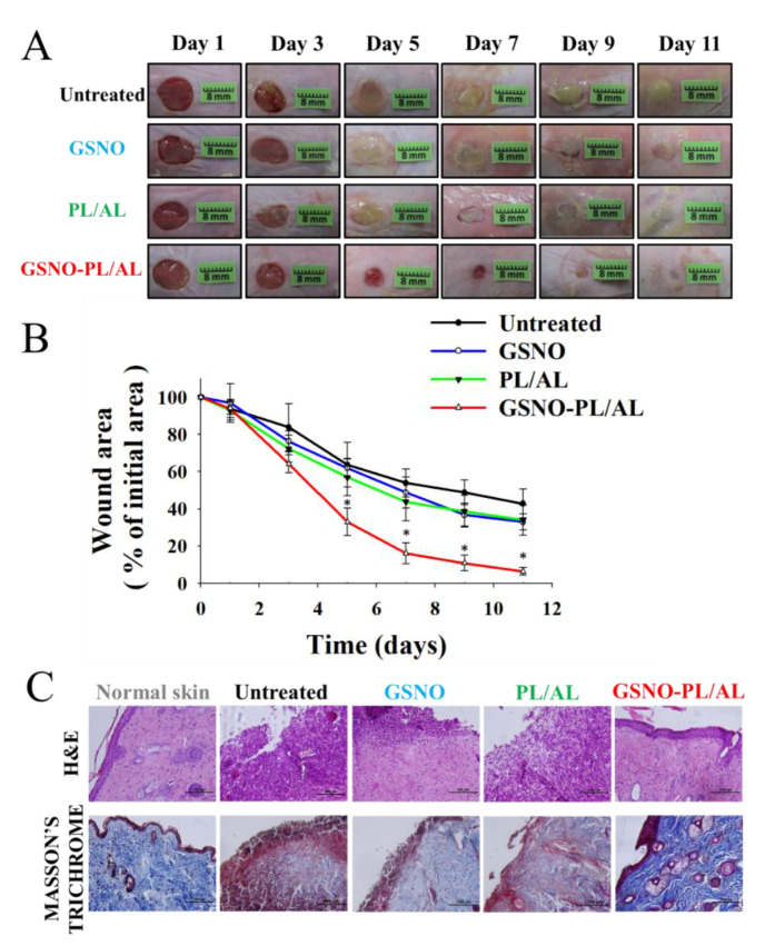
Figure 6.
(A) Representative photographs of healing burn wounds infected with MRPA in mice treated with PL/AL and GSNO-PL/AL. (B) Area reduction (%) profiles of MRPA-infected wounds. Data are presented as means ± SD; n = 6. * p < 0.05 vs. untreated. (C) Histological analysis (hematoxylin and eosin (H & E) and Masson’s trichrome staining) of MRPA-infected wound at 11 days after injury. Bar = 100 µm.

