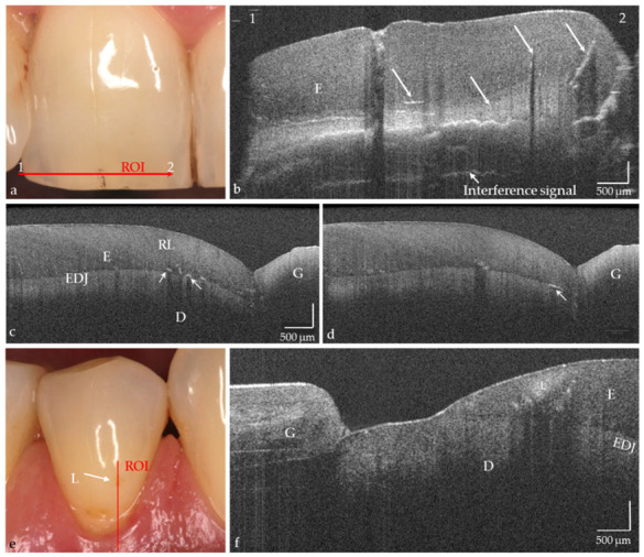
Figure 4.

In vivo imaging of hard tooth tissues (a–f), intrinsic structures, and defects in enamel ((b–d), arrows). Partially also Retzius lines (RL; (c)) and superficial tissue structures of the gingiva (f) are indicated. (e) Premolar with vestibular brown discoloration. The OCT cross-sectional image (f) showed that besides the discoloration a carious lesion in enamel (L) is present. The lesion body appears as a bright zone with clear shadowing. Scales are related to refractive index n = 1.0. While the horizontal scale in an OCT cross-sectional image is independent of the refractive index (n) of the tooth structures, the length of the vertical scale has to be divided by it (mean n for enamel and dentin approx. 1.5). Enamel (E), dentin (D), enamel-dentin junction (EDJ), gingiva (G), region of interest (ROI).
