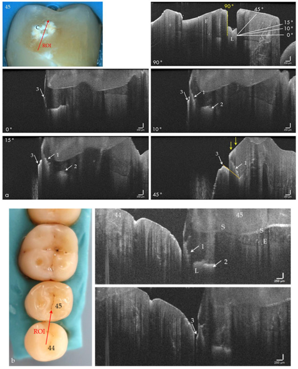
Figure 5.

(a) Extracted tooth 45 with a cavitated approximal enamel lesion (C). In the OCT image, recorded perpendicular (90°) to the surface, the lesion (L) appeared as a box-shaped structure in the enamel. If the cavitation is imaged from occlusal with an angle of incidence of 0° to 45°, the representation changes. The upper cavity wall appears as a diagonal signal line (arrows 1). Furthermore, the lesion body (arrows 2) and the cervical margin of the cavitation become visible (arrows 3). (b) Artificial row of teeth. In the ROI, the diagonal signal of the cavity (arrow 1) merges into a horizontal signal line representing the lesion body extending to the enamel-dentin junction (arrow 2). With a slight variation of the probe position, the protruding edge of the cavity entrance opening appears at some point (arrow 3). When focusing on deeper structures, the tooth surface (S) is inverted in the OCT image. The vertical scales are related to refractive index n = 1.0 (see the remark in Figure 4). Enamel (E).
