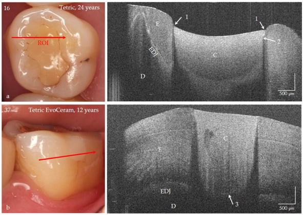
Figure 15.

Class I and V composite restorations (C) in teeth 16 and 37 after 24 and 12 years of clinical function, imaged with OCT from the occlusal (a) and vestibular view (b). (a) The surface of the composite restoration (Tetric, adhesive #) shows negative steps at the restoration margins (arrows 1). The marginal discoloration (*) can be explained by the short interfacial marginal gap and not by caries adjacent to the restoration margin (secondary caries) (arrow 2, bright line). Beyond that the bonding interface is still intact after 24 years (no bright signal lines) and the material is homogeneous. (b) At the floor of the 12-year-old restoration (Tetric EvoCeram, adhesive #) a short interfacial gap (3) is indicated, the progression of which can be monitored. Compared to tooth 16, the restoration margins are flat, and the material exhibits inhomogeneities. Enamel (E), dentin (D), the enamel-dentin junction (EDJ). The red arrows mark the section planes of the OCT cross-sectional images. The vertical scales are related to the refractive index n = 1.0 (see remark in Figure 4).
