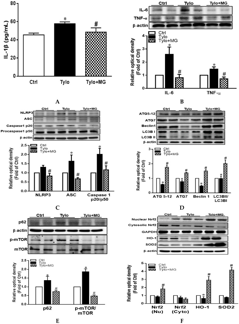
Figure 3.
Effects of magnolol (MG) treatment on interleukin-1β (IL-1β) secretion and NLR family pyrin domain-containing 3 (NLRP3) inflammasome activation in the liver of tyloxapol-injected rats. (A) Plasma levels of IL-1β; (B) representative Western blot and densitometry analysis of tumor necrosis factor-α (TNF-α) and interleukin-6 (IL-6); (C) representative Western blot and densitometry analysis of NLRP3, apoptosis-associated speck-like protein (ASC), and caspase 1 p20 and p50; (D) representative Western blot and densitometry analysis of autophagy related protein 5-12 (ATG5-12), ATG7, Beclin1, and microtubule-associated protein light chain 3 B II (LC3BII)/LC3BI ratio (E) representative Western blot and densitometry analysis of sequestosome-1 (SQSTM1/p62) and phosphorylation of mTOR; (F) representative Western blot and densitometry analysis of nuclear/cytosolic Nrf2 and the downstream HO-1 and SOD2. Data are expressed as mean ± SEM, n = 5–12. * p < 0.05 vs. Ctrl; # p < 0.05 vs. Tylo (tyloxapol).

