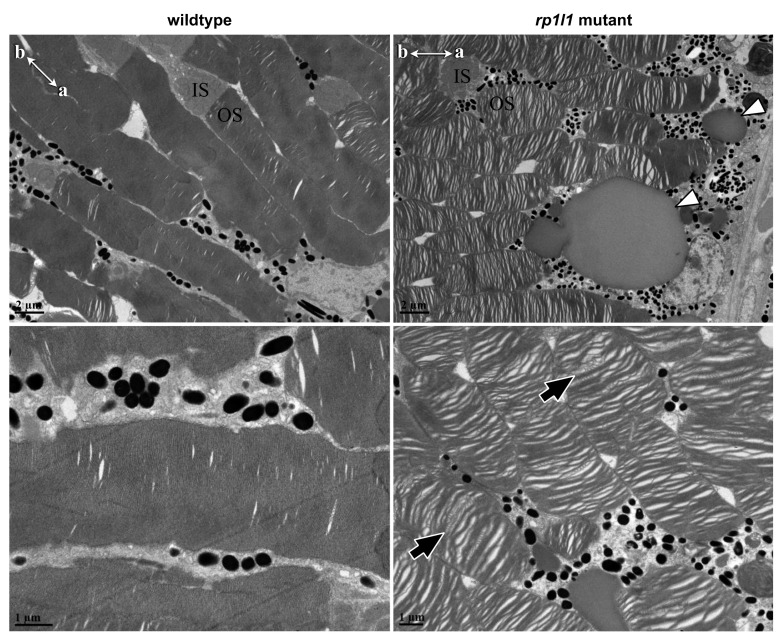
Figure 7.
rp1l1 mutant zebrafish have disorganized photoreceptor outer segments and deposits between the photoreceptors and retinal pigment epithelium. Electron microscopy of 11-month-old mutant and wild-type photoreceptors. Mutant photoreceptors appear disorganized, with gaps between the discs, and discs wave or swirl in some outer segments (black arrows). Additionally, there appear to be deposits between the photoreceptor outer segments and retinal pigment epithelium (white arrowheads). IS = inner segment; OS = outer segment. Compasses in top-left corners show apical (a) and basal (b) photoreceptor orientations for the images.

