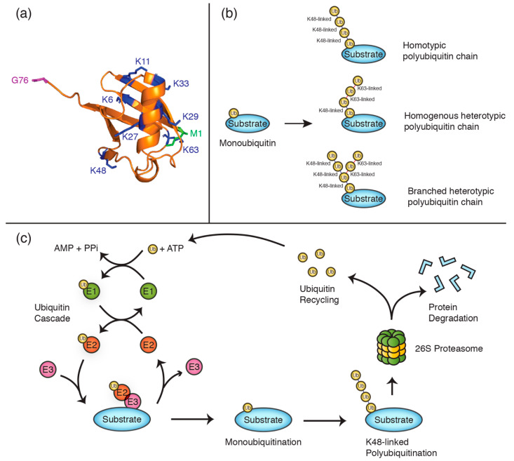
Figure 1.
Polyubiquitin chains are constituted by specific lysine linkages. (a) Cartoon structure of ubiquitin (PDB 1ubq) with lysine residues illustrated in blue, the N-terminal methionine (M1) in green, and the C-terminal glycine residue (G76) in purple. (b) Schematics exemplifying different polyubiquitin chain types. (c) Schematic of the ubiquitination cycle, illustrating the proteolytic degradation of a K48-linked polyubiquitinated substrate and ubiquitin recycling.

