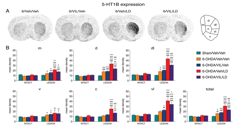
Figure 4.
Vilazodone inhibited the L-DOPA-induced increase in 5-HT1B expression in the dopamine-depleted striatum. (A) Illustrations of film autoradiograms depict expression of 5-HT1B in sections from the mid-level striatum in rats with a 6-OHDA lesion (right hemisphere) followed by repeated vehicle (6-OHDA/Veh/Veh), vilazodone (10 mg/kg) + vehicle (6-OHDA/VIL/Veh), vehicle + L-DOPA (5 mg/kg) (6-OHDA/Veh/LD), or vilazodone + L-DOPA treatment (6-OHDA/VIL/LD). Animals were killed 60 min after the L-DOPA injection. (B) Mean density values (mean ± SEM) for 5-HT1B expression in the striatum on the intact side or the side of the lesion in the sham lesion controls (Sham/Veh/Veh) and the 6-OHDA/Veh/Veh, 6-OHDA/VIL/Veh, 6-OHDA/Veh/LD, and 6-OHDA/VIL/LD groups, measured in the six sectors and the total middle striatum (upper right). m, medial; d, dorsal; dl, dorsolateral; vl, ventrolateral; c, central; v, ventral. # p < 0.05, ## p < 0.01, ### p < 0.001 vs. same group on the intact side; * p < 0.05, ** p < 0.01, *** p < 0.001 vs. Sham/Veh/Veh; φ p < 0.05, φφ p < 0.01, φφφ p < 0.001 vs. 6-OHDA/Veh/Veh; + p < 0.05, ++ p < 0.01, +++ p < 0.001 vs. 6-OHDA/VIL/Veh; $ p < 0.05, $$$ p < 0.001 vs. 6-OHDA/Veh/LD.

