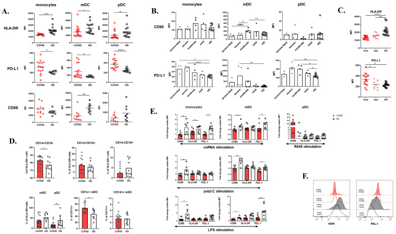
Figure 3.
Phenotype of monocytes and dendritic cells. (A) HLA-DR, PD-L1, and CD86 expression on monocytes, myeloid, and plasmacytoid dendritic cells (mDCs, pDCs) detected by flow cytometry. Data are expressed as mean fluorescence intensity (MFI). (B) CD80 and PD-L1 expression on monocytes, mDCs, and pDCs of COVID-19 (n = 15) patients divided according to the severity of the disease. Data are expressed as MFI. (C) HLA-DR and PD-L1 expression on COVID-19 monocytes detected after admission to the hospital (“first”, n = 17) and after time of 2–4 weeks (“last”, n = 9). Data are expressed as MFI. (D) Peripheral blood monocytes and DC subpopulations of COVID-19 patients (n = 14) and HD (n = 10) assessed by flow cytometry. (E) Whole blood was stimulated with 10 μg/mL ssRNA, 50μg/mL polyI:C, 1 μg/mL R848, 1 μg/mL LPS, or left untreated overnight and then CD86, HLA-DR, and PD-L1 expression on the surface of monocytes, mDCs, and pDCs were analyzed. Data are expressed as fold change (stimulated MFI/unstimulated MFI) of nine COVID-19 patients and eight HD. (F) Representative histograms of CD86 and PD-L1 expression upon ssRNA stimulation of COVID-19 patients and HD. Statistical analysis was performed using the Wilcoxon paired or Mann–Whitney unpaired t-test. Values of p < 0.05 (*), p < 0.01 (**), p < 0.001 (***), and p < 0.0001 (****) were considered statistically significant.

