Figure 1.
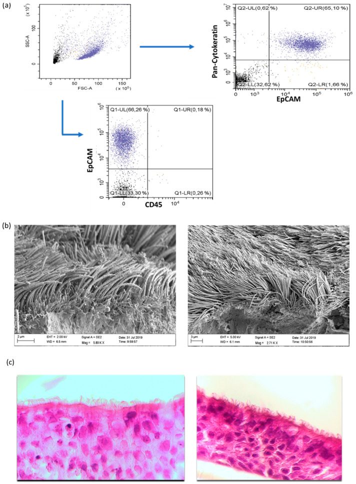
Characterization of primary human nasal epithelial cells (NAEPCs) derived from the pediatric exacerbation study cohort. (a) NAEPCs were obtained through nasal brushing procedure. After different washing and lysing steps, NAEPCs were seeded in collagen I pre-coated T75 flasks and cultured for up to 2 weeks until 80–90% confluency was reached. During the splitting process from passage 0 (P0) to passage 1 (P1), NAEPCs were harvested for flow cytometric analyses. Cells were stained with CD45-APC, CD326(EpCAM)-PE as surface antibodies. After fixation and permeabilization, anti-cytokeratin-FITC was used for intracellular staining. (b) After passage 2 (P2), the cells were seeded for organotypic 3D air–liquid interface cultures. Nasal mucus secretion and ciliary beats were observed through light microscopy after 6 to 8 weeks of culturing. Final specimens were then processed for raster electron microscopic imaging. (c) Histologic analyses confirmed the morphology of the nasal epithelial cell population (10× magnification).

