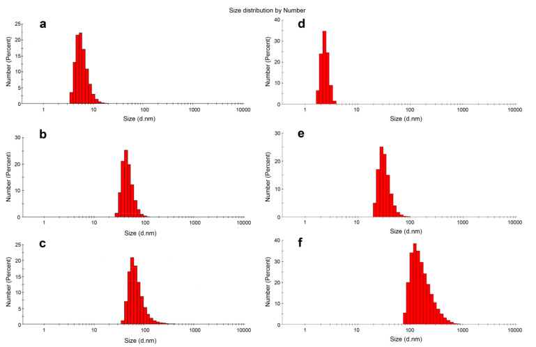
Figure 2.
Size distributions of the control oligonucleotides 13 (a), 17 (d) compared with D-13 (b), D-13PG (c), D-17PG (e) and D-17PG (f) conjugates (after 3 h incubation at 5 µM in TAM buffer) as measured by DLS. The D-13PG dodecyl oligonucleotide conjugate with two uncharged PG groups assembled into slightly enlarged micellar particles compared to its DNA analog D-13 (Table 1, Figure 2c). Interestingly, we observed a considerable increase in the size of D-17PG self-assemblies (Table 1, Figure 2f). We attributed these results to the multiple micellar complexes that appeared in a short time in this case, or to more complex micellar structures. Such micellar aggregates of 129.96 ± 73.36 (PDI 0.216) nm in diameter were detected after 24 h incubation for D-17 conjugate.

