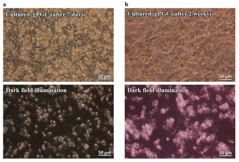
Figure 2.
The gPGCs cultured for 7 and 14 days, observed under an inverted phase-contrast microscope and in darkfield illumination. This technique allows us to clearly distinguish PGCs from the feeder layer and other cells. The PGCs are visible as bright, illuminated cells among the other cell types (P.434242 patent application number). Magnification with 40× objective (Leica DMI8).

