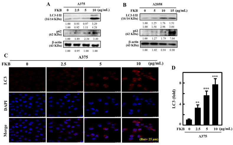
Figure 4.
FKB induced autophagy LC3-II and sequestosome 1 (p62) expression in human melanoma cells. (A) A375 and (B) A2058 cells were treated with various concentrations of FKB (0–10 or 0–15 μg/mL) for 24 h. These cells were subjected to Western blot analysis to determine the conversion of LC3-I to LC3-II and expression of p62 proteins. (C,D) FKB (0–10 μg/mL) was treated to A375 cells for 24 h. Immunofluorescence staining (100× magnification) was used to measure the accumulation of LC3. The data were expressed as fold-over untreated control cells. Each value was expressed as mean ± standard deviation (SD) of three experiments. Statistical significance was assigned as ** p < 0.01, and *** p < 0.001 as compared to untreated control cells.

