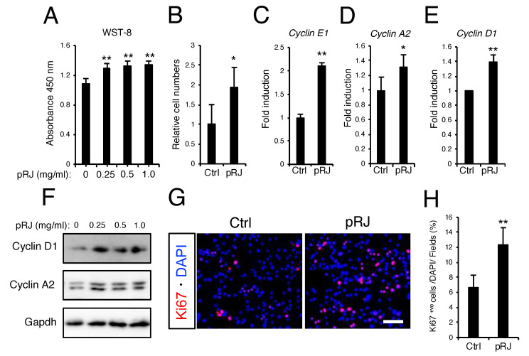
Figure 3.
pRJ stimulates proliferation of myoblasts in C2C12 cells. (A–B) C2C12 cells were treated with 0. 0.25, 0.5, or 1.0 mg/mL pRJ solution for 2 days. The number of living cells was assessed using Cell Counting kit-8. Cells were treated with 1.0 mg/mL pRJ for 2 days. After staining with trypan blue, the number of living cells was determined by direct counting. Graphs show the ratio of number of cells treated with pRJ divided by control (B). (C–E) The mRNA levels of indicated genes in cells treated with or without 1.0 mg/ml pRJ for 2 days. (F) Western blot showing protein levels of Cyclin D1, Cyclin A2, or Gapdh in C2C12 cells treated with 0. 0.25, 0.5, or 1.0 mg/mL pRJ solution for 2 days. (G and H) Images of Ki67 positive (+ve) immunostaining in cells treated with or without 1.0 mg/mL pRJ (G). The graph indicates the number of Ki67+ve cells as a percentage of total cells stained with DAPI (H). Images are representative of multiple independent experiments (F and G). Scale bar corresponds to 100 μm (G). Data are mean ± SD (n = 4). ∗∗p < 0.01, ∗p < 0.05, versus control (Ctrl).

